Laboratory Tests
KEYWORDS
for searching the Internet and other reference sources
Blood tests
Complete blood count (CBC)
Computerized tomography (CT) scan
Echocardiogram
ELISA
Erythrocyte sedimentation rote (ESR)
Gram stain
Laboratory culture
Magnetic resonance imaging (MRI)
Nucleic acid test (NAT)
Serology testing
Ultrasound
Urinalysis
X rays
What Are Laboratory Tests?
Before doctors can treat someone, they have to understand what is causing the illness. By talking with the patient ("taking the history"), examining the patient, and, when necessary, ordering tests (often called laboratory or lab tests), a doctor is better able to determine the illness. Most of the time, a doctor will be able to make the diagnosis without needing to order any tests. Thousands of possible tests exist, ranging from simple, inexpensive tests, such as looking at the color of urine, to much more complicated and expensive ones, such as certain types of X rays. Through training and experience, doctors learn when to order lab tests, which ones to order, and how to interpret the results.
Tests are meant to help identify the cause of a person's illness. Sometimes a test reveals the specific organism or cause and the diagnosis will be made relatively easily. For example, a person with a high fever, painful sore throat, and pus * on the tonsil * area may have a positive throat culture for Streptococcus (strep-tuh-KAH-kus) bacteria. The lab test, in this case a throat culture, confirms the diagnosis of strep throat.
Sometimes a lab test will only suggest the possibility of a specific cause and making the diagnosis will require much more judgment. For example, an elderly person feeling short of breath may have an X ray that could mean a type of heart disease, but more tests would be necessary to confirm that diagnosis.
* pus is a thick, creamy fluid, usually yellow or greenish in color, that forms at the site of an infection. Pus contains infection-fighting white cells and other substances.
A lab test also can eliminate a suspected cause, shortening the list of diagnoses to be considered. For example, a person with anemia * might have been tested for the amount of iron in the blood. A normal (or "negative") test would help eliminate the diagnosis of "iron-deficiency" anemia and help guide the evaluation toward other possibilities.
The History of Laboratory Testing
The existence of microbes has been known since Antonie van Leeuwenhoek first described "little animals in rain water" in 1674, but it was Robert Koch, the "father of modern bacteriology" (the study of bacteria), who proved that a specific organism caused a specific disease. His first discovery was with anthrax (AN-thraks), an infectious disease caused by the bacterium Bacillus anthracis. In 1876, Koch described the life cycle of the bacillus as it went from spore * to infectious agent. In 1881 Louis Pasteur took it one step further and was able to develop an effective vaccine against anthrax for sheep and cattle.
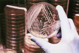
Following his work with anthrax, Koch set out to find a better way of growing and isolating microorganisms * . Previously, he had used liquid cultures, which were easily contaminated with other organisms, and he needed live animals as incubators to grow the bacteria he was studying. Based on his observations of colonies (groups visible to the naked eye) of microorganisms growing on a boiled potato, he set out to develop a solid substance (a nutrient medium) on which he could grow bacteria. With the help of his assistant Julius Richard Petri, who is remembered for the invention of the petri dish, he developed methods for isolating colonies of microorganisms.
The method for culturing bacteria that Koch developed over a century ago became the commonly accepted standard for use by labs. Since that time, culture methods have improved—and laboratories now also culture yeast, fungi * , and viruses * . How is that done? First, a sample of the infected fluid or tissue is obtained. This can be as simple as a swab of fluid taken from the throat or a urine sample, or it may be pus drained from an abscess * or a piece of tissue removed from an infected area (a procedure called a biopsy, BI-op-see). Then, under special conditions, the organisms are allowed to grow and are identified under the microscope. Additional testing may be done to determine whether the microorganism would be killed by various commonly used antibiotics * . This is how the doctor knows that the right antibiotic has been chosen to eliminate the organism. This step has become increasingly important because some bacteria that were once successfully treated with a certain antibiotic are now becoming resistant to it.
* tonsils are paired clusters of lymph tissues in the throat that help protect the body from bacteria and viruses that enter through a person's nose or mouth.
* anemia (uh-NEE-me-uh) is a blood condition in which there is a decreased amount of oxygen-carrying hemoglobin in the blood and, usually, fewer than normal numbers of red blood cells.
* spore is a temporarily inactive form of a germ enclosed in a protective shell.
* microorganisms are tiny organisms that can be seen only using a microscope. Types of microorganisms include fungi, bacteria, and viruses.
* fungi (FUNG-eye) are microorganisms that can grow in or on the body, causing infections of internal organs or of the skin, hair, and nails.
* viruses (VY-ruh-sez) are tiny infectious agents that can cause infectious diseases. A virus can only reproduce within the cells It infects.
* abscess (AB-ses) is a localized or walled-off accumulation of pus caused by infection that can occur anywhere in the body.
* antibiotics (an-tie-by-AH-tiks) are drugs that kill or slow the growth of bacteria.
* protozoa (pro-tuh-ZOH-uh) are single-celled microorganisms (tiny organisms), some of which are capable of causing disease in humans.
* trichomoniasis (trih-ko-mo-NYEuh-sis) is a common sexually transmitted disease caused by the parasite Trichomonas vaginalis.
Culture results usually take 24 to 48 hours, but some bacteria take a long time to grow, and those results may take a week or longer. In the meantime, doctors may use other tests that yield immediate results, such as looking at the organism under a microscope and staining techniques. These tests can help doctors make a diagnosis and decide on treatment while they are waiting for the final culture results. For example, the thrashing tail movements of the protozoa * Trichomonas vaginalis, the cause of trichomoniasis * , are easily seen on a slide under a microscope in the doctor's office. Staining (adding certain colors, or dyes) an organism allows the doctor to identify the organism based on its appearance. This is
Types of Laboratory Tests
Urinalysis
Urinalysis, or urine tests, allows for the rapid analysis of the urine. Special strips react by turning colors when dipped into a urine sample testing for blood, glucose (sugar), and other substances that may indicate infection. These "dipsticks" are a quick screen for possible urine infections, as well as other diseases such as diabetes * and problems with the kidneys. The urine also is examined under a microscope to look for bacteria, and the urine sample is sent to the lab to be cultured for bacteria if a urinary infection is suspected.
Stool tests
Just as doctors can examine and test the urine, they are able to examine the stool by a number of methods. Doctors can test for the presence of illness-causing bacteria, pus, blood, or unusual chemicals. Stool can be cultured for the bacteria that may cause diseases such as typhoid fever. The color of the stool may give evidence as to the cause of the problem. For example, very light stools could suggest a type of hepatitis.
* meningitis (meh-nin-JY-tis) is an inflammation of the meninges, the membranes that surround the brain and the spinal cord. Meningitis is most often caused by infection with a virus or a bacterium.
* diabetes (dye-uh-BEE-teez) is a condition in which the body's pancreas does not produce enough insulin or the body cannot use the insulin it makes effectively, resulting in increased levels of sugar in the blood. This can lead to increased urination, dehydration, weight loss, weakness, and a number of other symptoms and complications related to chemical imbalances within the body.
Blood tests
Blood tests may be used to look for general signs of infection or to diagnose a specific infection. A complete blood count (CBC) measures the different parts that make up blood, including the numbers of red blood cells, white blood cells, and platelets * ; the total amount of hemoglobin * in the blood; and even the size of red blood cells. During a bacterial infection, the number of white cells is often increased because they help fight off infection. If a person has anemia, this suggests the infection is attacking the red blood cells, as is seen with malaria * . A long-standing infection may also cause anemia. The platelets are involved in clotting * the blood, and their numbers may drop in some serious and overwhelming infections, such as meningitis. A CBC screen often is ordered when an infectious disease is suspected. Although it is not used to diagnose the cause, it is helpful for seeing how the body is reacting to an infection.
Spinal tap (lumbar puncture)
Sometimes, when an infection involving the brain or the coverings of the brain is suspected, doctors need to test the fluid that surrounds the brain. That type of infection, known as meningitis (the coverings of the brain are the meninges), can be life-threatening—and needs to be diagnosed and treated right away. One of the ways doctors can make the diagnosis is by a spinal tap (also known as a lumbar puncture or LP). This occurs when a doctor places a very thin hollow tube through a space between the bones (the vertebrae) in the lower back and then takes out a small amount of spinal fluid. Spinal fluid is the liquid that surrounds the brain and the spinal cord. By testing spinal fluid for bacteria and other substances, doctors can tell whether an infection is the cause of the person's illness.
Antibody and antigen tests
Antibody tests are commonly used to diagnose many infectious diseases. These tests measure the body's response to an infectious disease. Antibodies * are produced by the immune system * in response to antigens * found on the surface of the invading organism. It takes some time for the body to mount an antibody response to a new infection, so this test may not be useful when an illness is starting. The test is often repeated weeks later to measure any increase in antibodies. Once they are formed, many antibodies are present long after the infection is gone. This often (but not always) gives a person immunity, or protection, from future infection. Many infections are diagnosed using antibody testing, including hepatitis, Lyme disease, and HIV infection.
Unlike antibody tests (which indirectly measure the body's response to an infection), antigen tests directly detect the presence of the organism by identifying the specific proteins that the infectious organism has on its surface. Unlike antibody tests (which might indicate that there was an infection in the past), the presence of antigens most likely means the infection is present at that moment. Examples of antigen testing include rapid strep tests, chlamydia * tests, and some HIV testing.
* platelets (PLATE-lets) are tiny disk-shaped particles within the blood that play an important role in clotting.
* hemoglobin (HE-muh-glo-bin) is the oxygen-carrying pigment of the red blood cells.
* malaria (mah-LAIR-e-uh) is a disease spread to humans by the bite of an infected mosquito.
* clotting is the body's way of thickening blood to stop bleeding.
* antibodies (AN-tih-bah-deez) are protein molecules produced by the body's immune system to help fight specific infections caused by microorganisms, such as bacteria and viruses.
* immune system is the system of the body composed of specialized cells and the substances they produce that helps protect the body against disease-causing germs.
* antigens (AN-tih-jenz) are substances that are recognized as a threat by the body's immune system, which triggers the formation of specific antibodies against the substances.
* chlamydio (kla-MIH-dee-uh) are microorganisms in the Chlamydia family that can infect the urinary tract, genitals, eye, and respiratory tract, including the lungs.
* DNA, or deoxyribonucleic acid (dee-OX-see-ry-bo-nyoo-klay-ik AH-sid), is the specialized chemical substance that contains the genetic code necessary to build and maintain the structures and functions of living organisms.
Nucleic acid tests
Nucleic acid tests (NATs) are methods to identify tiny amounts of infectious organisms by multiplying (or amplifying) a small amount of that organism's DNA * to create a larger sample that can then be analyzed. They are a new and valuable tool in the identification of infectious agents, particularly those that are difficult to culture. NATs are currently used in the diagnosis of hepatitis C and HIV and are useful in making the diagnosis in infants because antibody testing is not reliable at such a young age. NATs have also been used in the diagnosis of Helicobacter pylori * infection, and tests are being developed for the rapid diagnosis of many serious infectious diseases.
Imaging tests
Imaging tests, using a variety of technologies, give physicians pictures of the inside of the body.
X RAYS
The use of X rays helps physicians "see" into the body without opening it. In general, X rays involve aiming a controlled amount of radiation at a specific area of the body. When the radiation passes through that part, it exposes a sort of photographic plate on the other side. If the part of the body that the radiation passes through is dense (like bone) or has a high water content (for example, lungs infected with pneumonia * ), the image on the plate will appear whiter. On occasion, doctors use a special liquid to improve the contrast between light and dark, which helps them better distinguish between different organs or see things in greater detail. This contrast dye may be injected into veins or swallowed.
BONE SCANS
A bone scan is a type of X ray that helps doctors locate areas of infection or cancer deep within the bone. It does this by revealing spots of increased or decreased bone cell activity. First, a radiotracer * , which can illuminate certain areas when scanned, is injected into a vein. The scan is performed hours later, once the radiotracer has had time to circulate in the body. A computer records the data from the scan and translates it into an image. By comparing places on the image where the tracer has (or has not) collected, doctors can pick out problem areas where bones may be damaged or infected.
COMPUTERIZED TOMOGRAPHY
Computerized tomography (kom-PYOO-terized toe-MAH-gruh-fee), or CT, scans X ray the body from a variety of angles. A scanner detects the X ray beams and transmits those data to a computer, which shapes the data into a series of images or photographs.
MAGNETIC RESONANCE IMAGING
Magnetic resonance imaging (MRI) uses a super-strong magnetic field, rather than X rays, to help create images. The magnetic field causes the protons in the body's water to vibrate a certain way, and the MRI machine records those vibrations and develops images based on them.
* Helicobacter pylori (HEEL-ih-kobak-ter pie-LOR-eye) is a bacterium that causes inflammation and ulcers, or sores, in the lining of the stomach and the upper part of the small intestine, also known as peptic ulcer disease.
* pneumonia (nu-MO-nyah) is inflammation of the lung.
* radiotracer is a substance that contains radioactive material.
ULTRASOUND
In ultrasound, ultra-high-frequency waves are beamed into the body, where they bounce off various structures. Somewhat similar in principle to sonar in submarines, ultrasound is painless and cannot be heard by the human ear. The machine records where the waves strike and bounce back and interprets this data, creating images. Ultrasound is widely used to help make diagnoses. One of the most common uses is part of regular prenatal care, when ultrasound is used to look at a baby in the womb to make sure it is developing normally. Ultrasound can be used to check specific organs, such as the liver * or kidneys, to look for unusual masses (tumors) or for abnormal size or density, such as might be seen with an abscess. An ultrasound image may appear as a single image, somewhat like a photograph, or as a moving image, like a video or movie.
ECHOCARDIOGRAM
An echocardiogram (eh-ko-KAR-dee-uh-gram) is a specific type of ultrasound that sends sound waves into the chest to "paint a picture" of the heart's structure. This test can be used to see the size of the heart's valves and chambers, how well they move, and other qualities that a physician would need to know.
Other tests
Other techniques also are used to identify infections. Some are very complex and expensive. Others are simple and cost almost nothing. For example, smell can sometimes help identify certain bacteria. Pseudomonas (which can cause urinary tract * infections and ear infections) smells sweet and fruity. The "whiff test" is a technique for diagnosing bacterial vaginosis, an overgrowth of vaginal bacteria. The chemical potassium hydroxide (KOH) is added to a small sample of fluid from the vagina. If the sample contains large numbers of bacteria, the KOH will make it smell fishy. A doctor also might add a few drops of KOH to a slide with scrapings from a suspected fungal infection, which will highlight the fungus. Using an ultraviolet or black light is another easy way to diagnose some fungal infections: the infected area glows when the light shines on it. Once the diagnosis of bacterial vaginosis is made, it is easily treated by a doctor.
* liver is a large organ located beneath the ribs on the right side of the body. The liver performs numerous digestive and chemical functions essential for health.
* urinary tract (YOOR-ih-nair-e TRAKT) is the system of organs and channels that makes urine and removes it from the body. It consists of the urethra, bladder, ureters, and kidneys.
Resources
Organization
U.S. National Library of Medicine, 8600 Rockville Pike, Bethesda, MD
20894. The National Library of Medicine has a website packed with
information on diseases and drugs, consumer resources, dictionaries and
encyclopedias of medical terms, and directories of doctors and helpful
organizations.
Telephone 888-346-3656
http://www.nlm.nih.gov
Website
KidsHealth.org
. KidsHealth is a website created by the medical experts of the Nemours
Foundation and is devoted to issues of children's health. It
contains articles on a variety of health topics, including why doctors
order laboratory tests.
http://www.KidsHealth.org
Comment about this article, ask questions, or add new information about this topic: