Spina Bifida
Spina bifida (SPY-na BI-fi-da) is a birth defect in which the spinal column does not form properly, leaving a gap or opening in the spine.
KEYWORDS
for searching the Internet and other reference sources
Meningocele
Myeomeningocele
Neural tube defects
Neuology
Orthopedics
Brian Teaches Class
As part of a sixth grade science project, Brian chose to report on a condition called spina bifida. He showed a picture of the ring-shaped bones, or vertebrae, of the spine and demonstrated how the vertebrae protect the spinal cord and anchor muscles. He explained that in people with spina bifida, some of the bony plates that should cover the spine do not close, leaving an unprotected opening at the back of the spine.
No one in Brian's class had ever heard of spina bifida, and they all were surprised to learn that Brian had been born with it. He had a mild form of the condition that was corrected surgically when he was an infant. He ended his presentation by showing the small scar on his lower back.
What Is Spina Bifida?
Spina bifida is a Latin term meaning "split spine" or open spine." It is the most common of several birth defects called neural tube defects. The neural tube contains the cells that ultimately make the spinal cord and the brain, and it develops during the first three to four weeks of pregnancy (often before a woman even knows that she is pregnant).
Spina bifida results when the sides of the neural tube fail to join together properly, leaving an open area. Often the gap occurs in the lower back at the base of the spine. The spinal cord is part of the central nervous system, which allows a person to move and sense the world around them. Thus, because spina bifida involves the central nervous system, it can cause a range of physical and mental problems.
Prenatal Testing for Spina Bifida
Sometimes parents can find out whether their baby has spina bifida before the baby is born. There are several commonly used tests.
The maternal-serum alfa-fetoprotein (AFP) test is performed between the sixteenth and eighteenth weeks of pregnancy. Alfa-fetoprotein is a substance made by the developing fetus. Because the mother and fetus are connected via their circulatory systems, AFP from the fetus gets into the mother's bloodstream. By measuring the amount of AFP in the mother's blood, doctors get an indication of the likelihood that the fetus has certain birth defects. This test does not give a definite answer, and high levels of AFP only suggest that the fetus might have spina bifida. If AFP levels are high, the test is repeated. If still high, other tests are needed to confirm that the fetus has spina bifida. Many times, high AFP readings are false alarms and the baby is just fine.
Ultrasound can be used to confirm or rule out spina bifida. An ultrasound works by bouncing sound waves off of internal structures. A computer converts the returning sound waves into an image of the fetus inside the uterus. Sometimes the defect in the developing spine is visible on the ultrasound image.
Amniocentesis is a procedure performed between the sixteenth and eighteenth week of pregnancy. In this procedure, a needle is passed through the mother's belly into her uterus to collect some of the fluid in which the fetus lives. This fluid, called amniotic fluid, contains cells and chemicals from the fetus. Levels of AFP can be measured to determine whether the fetus has spina bifida.
Is Spina Bifida Always Serious?
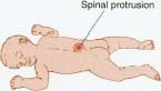
Spina bifida is a common birth defect, but it does not always cause serious problems. At birth, the gap may be so slight that it is invisible and harmless. On the other hand, sometimes the spinal cord bulges out through the mal-formed vertebrae and there are serious neurological (nerve) problems.
Spina bifida occulta
Brian was born with spina bifida occulta, the mildest form of spina bifida. "Occulta" means hidden, and in many cases, the gap in the spine is never detected. Often there is an opening in one or two of the vertebrae but the spinal cord is not affected. A dimple, a birthmark, or a patch of hair may be visible on the skin overlying the site of the gap.
Scientists estimate that about 40 percent of all Americans have this form of spina bifida, but not many ever know they have it. Most people with spina bifida occulta never need treatment. Brian was an exception. He needed surgery because as he grew, the lower end of his spinal cord got caught against his vertebrae, causing him to have problems controlling his bladder. The doctors "unhooked" the spinal cord and closed the gap surgically.
Spina bifida manifesta
Spina bifida manifesta includes two forms of spina bifida that together represent one of the most common disabling birth defects. On average, 1 out of 1,000 babies in the United States is born with either meningocele (me-NING-go-seel) or myelomeningocele (MY-e-lo-me-NING-go-seel).
Meningocele
Of babies born with spina bifida manifesta, about 4 percent have the meningocele form. The meninges (me-NIN-jez) consist of three layers of tough membranes that cover and protect the brain and spinal cord. The brain and spinal cord also are bathed in a fluid called cerebro-spinal fluid (CSF). A meningocele is a CSF-filled sac formed when the meninges balloon through the gap in the vertebrae. It looks like a large blister covered by a thin layer of skin. The sac can range in size from as small as a grape to as large as a grapefruit.
A meningocele is harmless if the sac contains only CSF. However, if nerves are caught in the sac, the affected baby can have problems controlling muscles and the bladder. Babies with this form of spina bifida usually have surgery during infancy to put the meninges back inside the vertebrae and to close the gap in the vertebrae.
Myelomeningocele
When most people think of spina bifida, they think of the myelomeningocele form. Approximately 96 percent of babies born with spina bifida manifesta have myelomeningocele, and it is the most serious type of spina bifida. As in meningocele, the meninges bulge out through the gap in the spine, but in myelomeningocele, part of the spinal cord bulges out as well. The sac can be covered with skin or the nerves can actually be exposed.
People with myelomeningocele have a variety of physical and mental problems, the severity of which depends on where the defect in the spine occurs. A gap high up on the spinal column will create more problems than a gap at the lower back. People with this condition usually experience loss of movement and feeling (paralysis) below the abnormal verte-brae. The most severely impaired children cannot walk or control their bowel or bladder. Most babies born with myelomeningocele also have hydrocephalus, which means that they have too much fluid inside and surrounding their brain. If hydrocephalus is not treated, the excess pressure in the skull can cause blindness and permanent brain damage.
Myelomeningocele requires surgery within 24 to 48 hours of birth. Surgeons must close the gap in the vertebrae to protect the spinal cord and prevent infection. They also must treat hydrocephalus, if present. They do this by placing a device called a shunt into the brain to drain excess fluid and relieve pressure on the brain.
What Causes Spina Bifida?
Spina bifida sometimes runs in families, which suggests that genes may play a role in some cases, but 90 to 95 percent of babies with spina bifida are born to families that have never before had a child with the condition. Mothers who have diabetes, a high fever during pregnancy, or who have taken a drug called valproic acid to treat epilepsy seem to have a greater chance of having a baby with spina bifida than mothers without these conditions. There also appears to be a link between a deficiency of folic acid (a B vitamin) in the mother's diet and having a baby with spina bifida. Adding folic acid to the diet significantly reduces the chance that a baby will be born with spina bifida.
Living with Spina Bifida
Most children with spina bifida occulta, and many with meningocele, live normal lives without any impairment. Children born with myelomeningocele, however, often have multiple problems resulting from damage to their spinal cord. Surgery to repair the gap in the vertebrae and to place a shunt in the brain can prevent further damage to the nervous system. However, it cannot reverse the nerve problems that already are present at birth.
The severity of symptoms caused by myelomeningocele varies from child to child. Common problems, however, include the inability to control the bowel and bladder. Catheters * , diapers, and attentive care-givers can help alleviate embarrassment caused by these problems.
Preventing Spina Bifida:
The Role of Folic Acid
Scientists estimate that the incidence of spina bifida can be decreased by as much as 75 percent if all women of child-bearing age consume 0.4 mg of folic acid each day.
Spina bifida has been linked to a deficiency of folic acid during the first weeks of pregnancy. Folic acid is one of the B vitamins and it is essential for proper functioning of the human body. When the body is growing quickly, such as during pregnancy and during fetal development, the body needs more folic acid than usual.
Good sources of folic acid include dark green leafy vegetables (like spinach and broccoli), eggs, and orange juice. In addition, the U.S. Food and Drug Administration requires breads and enriched grains and cereals to have folic acid added to them. Even with folic acid supplements added to common foods, the average American diet does not contain 0.4 mg of folic acid per day. Most multi-vitamins, however, now contain the recommended dose of folic acid.
* catheters (KATH-e-terz) are tubes inserted into parts of the body to allow fluids to flow in and out. Catheters into the bladder through the urethra (the last part of the urinary tract) allow urine to flow into an outside container.
Many affected children cannot walk without crutches or leg braces, and many need a wheelchair. In addition, some children have learning problems, particularly with reading and math. For children with these problems, special education classes can help to prepare them for school.
Children with spina bifida often develop sensitivity or an allergy to latex (natural rubber), which is used in such healthcare products as gloves and catheter tubes, probably because they come into contact with latex so often and from such a young age.
Even with the disabilities caused by spina bifida, children with the disease often live well into adulthood. With the help of early and continuing medical, psychological, and educational treatment, children with spina bifida can lead full and productive lives.
See also
Birth Defects
Hydrocephalus
Incontinence
Paralysis
Resources
Books
Sandler, Adrian. Living with Spina Bifida: A Guide for Families and Professionals. Chapel Hill: University of North Carolina Press, 1997.
Lutkenhoff, Marlene. Spinabilities: A Young Person's Guide to Spina Bifida. Bethesda: Woodbine House, 1997.
Organizations
Spina Bifida Association of America, 4590 MacArthur Boulevard, Suite
250, Washington, DC 20007.
Telephone 800-621-3141
http://www.sbaa.org
Association for Spina Bifida and Hydrocephalus, 42 Park Road,
Peterborough, PE1 2UQ, England.
Telephone 01733-555988
http://www.asbah.demon.co.uk
March of Dimes Foundation, 1275 Mamaroneck Avenue, White Plains, NY
10605.
Telephone 888-663-4637
http://www.modimes.org/
Comment about this article, ask questions, or add new information about this topic: